产品说明
一般描述
Lymphocyte activation gene 3 protein (UniProt Q61790; also known as CD223, LAG-3) is encoded by the Lag3 gene (Gene ID 16768) in murine species. LAG-3 is an inhibitory T-cell surface molecule that modulates T-cell activation and homeostasis. LAG-3 co-localizes with CD8 and CD4 upon TCR engagement and alters TCR signaling. LAG-3 suppresses homeostasis proliferation (HP) of both lymphocytes and some dendritic cell populations in vivo, and enhanced HP is observed in mice deficient in LAG-3. LAG-3 signaling is shown to negatively regulate STAT5 phosphorylation in activated T cells, and no enhancement of HP was seen upon LAG-3 blockade in mice deficient in both STAT5a and STAT5b. Murine LAG-3 is produced with a signal peptide sequence (a.a. 1-22), the removal of which yields the mature protein with a large extracellular region (a.a. 23-442), followed by a transmembrane segment (a.a. 443-463) and a cytoplasmic tail (a.a. 464-521). The extracellular portion contains a V-type Ig-like domain (a.a. 37-163), followed by three C2-type Ig-like domains (a.a. 165-246, 258-341, and 345-412) and elven tandem repeats of 2-amino acid E-X seqeunce at its C-terminal end (a.a. 493-518).
特异性
Clone 4-10-C9 immunostained surface LAG-3 on activated CD4+ T cells by targeting an extracellular epitope within the third and fourth Ig-like domains (D3/D4 domains; second and third C2-type Ig-like domains). Surface LAG-3 degradation by pronase treatment abolished cell surface staining (Woo, S.R., et al. (2010). Eur. J. Immunol. 40(6):1768-1777).
免疫原
Murine LAG-3-expressing mouse T-cell hybridoma.
Epitope: Within D3/D4 domains.
应用
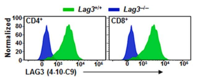
Flow Cytometry Analysis: 2.5 µg/mL from a representative lot detected surface LAG-3 immunoreactivity among the CD4+ and CD8+ populations of wild-type, but not Lag3-knockout, mouse splenocytes activated in vitro via CD3 cross-linking (Courtesy of Dario A. Vignali, Ph.D., University of Pittsburgh, PA, U.S.A.).
Flow Cytometry Analysis: A representative lot was fluorescently conjugated and detected an increased number of LAG-3-positive cells within the CD4+ and CD8+ populations of infiltrating lymphocytes (TILs) in tumors developed in mice exografted with murine B16 melanoma, MC38 colon adenocarcinoma, or Sa1N fibrosarcoma cells (Woo, S.R., et al. (2012). Cancer Res. 72(4): 917–927).
Flow Cytometry Analysis: A representative lot, pre-conjugated with Alexa Fluor® 647, detected both surface and intracellular LAG-3 by immunofluorescent staining of non-permeabilized and permeabilized primary murine CD4+ T cells activated in vitro via CD3 & CD28 cross-linking by immobilized antibodies. Pronase treatment of cells prior to permeabilization abolished cell surface staining (Woo, S.R., et al. (2010). Eur. J. Immunol. 40(6):1768-1777).
Flow Cytometry Analysis: A representative lot, pre-conjugated with Alexa Fluor
Immunocytochemistry Analysis: A representative lot detected both surface and intracellular LAG-3 by fluorescent immunocytochemistry staining of non-permeabilized and permeabilized primary murine CD4+ T cells activated in vitro via CD3 & CD28 cross-linking by immobilized antibodies. Pronase treatment of cells prior to permeabilization abolished cell surface staining (Woo, S.R., et al. (2010). Eur. J. Immunol. 40(6):1768-1777).
Immunocytochemistry Analysis: A representative lot detected intracellular LAG-3 immunoreactivity co-localized with those of the early and recycling endosome marker EEA1, as well as endosomal markers Rab11b and Rab27a by fluorescent immunocytochemistry staining of activated murine CD4+ T cells following pronase treatment and permeabilization (Woo, S.R., et al. (2010). Eur. J. Immunol. 40(6):1768-1777).
This Anti-LAG3 Antibody, clone 4-10-C9 is validated for use in Flow Cytometry, Immunocytochemistry for the detection of LAG3.
质量
Evaluated by Flow Cytometry in mouse splenocytes.
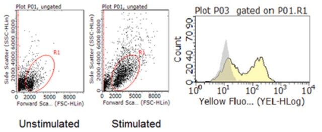
Flow Cytometry Analysis: 1 µg/mL of this antibody detected an induction of LAG-3-positive population in isolated mouse splenocytes following a 3-day 2 µg/mL Concanavalin A (Con A) stimulation.
目标描述
54.51/56.98 kDa (mature/pro-form) calculated.
外形
Protein G purified.
Format: Purified
其他说明
Concentration: Please refer to lot specific datasheet.
法律信息
ALEXA FLUOR is a registered trademark of Life Technologies
免责声明
Unless otherwise stated in our catalog or other company documentation accompanying the product(s), our products are intended for research use only and are not to be used for any other purpose, which includes but is not limited to, unauthorized commercial uses, in vitro diagnostic uses, ex vivo or in vivo therapeutic uses or any type of consumption or application to humans or animals.
基本信息
| eCl@ss | 32160702 |
| NACRES | NA.41 |
产品性质
| 质量水平 | 100 |
| 生物来源 | mouse |
| 抗体形式 | purified immunoglobulin |
| antibody product type | primary antibodies |
| 克隆 | 4-10-C9, monoclonal |
| species reactivity | mouse |
| technique(s) | flow cytometry: suitable immunocytochemistry: suitable |
| 同位素/亚型 | IgG2aκ |
| NCBI登记号 | NP_032505 |
| UniProt登记号 | Q61790 |
| 运输 | wet ice |
安全信息
| 储存分类代码 | 12 - Non Combustible Liquids |
| WGK | WGK 1 |
| 闪点(F) | Not applicable |
| 闪点(C) | Not applicable |
Sigma-Aldrich

 m.cnreagent.com