
产品说明
一般描述
MLKL, also known as Mixed lineage kinase domain-like protein, and encoded by the gene MLKL, is an interesting protein that is required for the execution of programmed necrosis or necroptosis, for example in response to TNF-alpha induced necrosis. MLKL is a component of the "necrosome," the multiprotein complex that triggers tumor necrosis factor (TNF)-induced cell death by necroptosis. The programmed necrosis induced by TNF-α also requires the activities of the receptor-interacting serine-threonine kinases RIP1 and RIP3 and they too interact directly with the mixed lineage kinase domain-like protein MLKL. RIP3 is an essential upstream kinase in necroptosis. The pseudokinase MLKL functions as a substrate of RIP3 to mediate downstream signaling and when complexed to MLKL the RIP3-MLKL complex is stabilized by AMP-PNP to adopt an inactive conformation.
免疫原
Epitope: Brace region.
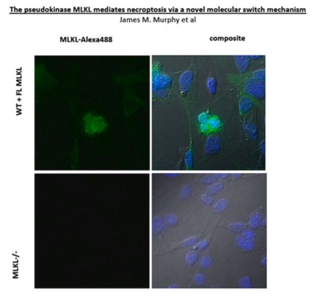
KLH-conjugated linear peptide corresponding to a sequence from the two-helix linker (Brace) region N-terminal to the pseudokinase domain of mouse MLKL (Hildebrand, J.M., et al. (2014). Proc. Natl. Acad. Sci. U.S.A. 111(42):15072-15077; Murphy, J.M., et al. (2013). Immunity. 39(3):443-453).
应用
This Anti-MLKL Antibody, clone 3H1 is validated for use in Immunocytochemistry, Immunoprecipitation, and Western Blotting for the detection of MLKL.
Immunocytochemistry Analysis: A representative lot detected MLKL in mouse dermal fibroblasts (MDFs) from wild-type, but not Mlkl-/- mice (Courtesy of Prof. James M. Murphy, Walter and Eliza Hall Institute, Australia).
Immunoprecipitation Analysis: A representative lot immunoprecipitated MLKL from soluble extract of Mlkl-/- mouse dermal fibroblasts (MDFs) harboring doxycycline-inducible wild-type mouse MLKL expression construct only after, but not before doxycycline treatment (Murphy, J.M., et al. (2013). Immunity. 39(3):443-453).
Western Blotting Analysis: A representative lot detected Q-VD-OPh (TSQ; Cat. No. 551476) treatment-induced membrane translocation of murine and equine MLKL N-terminal fragment (a.a. 1-180 and 1-189, respectively) exogenously expressed in mouse dermal fibroblasts (MDFs) from Mlkl-/- mice (Tanzer, M.C., et al. (2016). Cell Death Differ.. In press).
Western Blotting Analysis: A representative lot detected MLKL in human HT-29 and U937 cells (Tanzer, M.C., et al. (2015). Biochem. J. 471(2):255-265).
Western Blotting Analysis: Representative lots detected MLKL membrane translocation in mouse dermal fibroblasts (MDFs) upon Q-VD-OPh (TSQ; Cat. No. 551476) treatment (Tanzer, M.C., et al. (2015). Biochem. J. 471(2):255-265; Hildebrand, J.M., et al. (2014). Proc. Natl. Acad. Sci. U.S.A. 111(42):15072-15077).
Western Blotting Analysis: Representative lots detected MLKL expression in L292 mouse fibroblasts, as well as in mouse dermal fibroblasts (MDFs), mouse embryonic fibroblasts (MEFs) and bone marrow derived macrophages (BMDMs) from wild-type, but not Mlkl-/- mice (Cook, W.D., et al. (2014). Cell Death Differ. 21(10):1600-1612; Murphy, J.M., et al. (2013). Immunity. 39(3):443-453).
Western Blotting Analysis: A representative lot detected recombinant full-length mouse MLKL as well as a.a. 1-180 and a.a. 124-464, but not a.a. 1-125 or a.a. 179-464, MLKL fragments (Hildebrand, J.M., et al. (2014). Proc. Natl. Acad. Sci. U.S.A. 111(42):15072-15077).
Western Blotting Analysis: A representative lot detected MLKL expression in all tissues tested except brain from wild-type mice, while no target band was seen in any tissues from Mlkl-/- mice (Murphy, J.M., et al. (2013). Immunity. 39(3):443-453).
质量
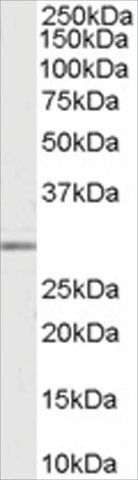
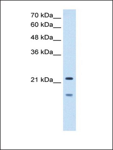
Evaluated by Western Blotting in mouse heart tissue lysate.
Western Blotting Analysis: 0.5 µg/mL of this antibody detected MLKL in 10 µg of mouse heart tissue lysate.
目标描述
~52 kDa observed. 54.48/30.28 (human isoform 1/2) and 54.32/53.38 kDa (mouse isoform 1/2) calculated. Uncharacterized band(s) may be observed in some cell lysates.
外形
Protein G purified.
Purified rat IgG in buffer containing 0.1 M Tris-Glycine (pH 7.4), 150 mM NaCl with 0.05% sodium azide.
Format: Purified
其他说明
Concentration: Please refer to the Certificate of Analysis for the lot-specific concentration.
免责声明
Unless otherwise stated in our catalog or other company documentation accompanying the product(s), our products are intended for research use only and are not to be used for any other purpose, which includes but is not limited to, unauthorized commercial uses, in vitro diagnostic uses, ex vivo or in vivo therapeutic uses or any type of consumption or application to humans or animals.
基本信息
| eCl@ss | 32160702 |
| NACRES | NA.41 |
产品性质
| 质量水平 | 100 |
| 生物来源 | rat |
| 抗体形式 | purified antibody |
| antibody product type | primary antibodies |
| 克隆 | 3H1, monoclonal |
| species reactivity | mouse |
| technique(s) | immunocytochemistry: suitable immunoprecipitation (IP): suitable western blot: suitable |
| 同位素/亚型 | IgG |
| NCBI登记号 | NP_083281 |
| UniProt登记号 | Q9D2Y4 |
| 运输 | wet ice |
安全信息
| 储存分类代码 | 12 - Non Combustible Liquids |
| WGK | WGK 1 |
| 闪点(F) | Not applicable |
| 闪点(C) | Not applicable |
Sigma-Aldrich

 m.cnreagent.com