产品说明
一般描述
Synapsin-1 (UniProt P17599; also known as Synapsin I) is encoded by the SYN1 gene (Gene ID 281510) in bovine species. The synapins constitute a family of abundant neuronal phosphoproteins that regulate SV trafficking and neurotransmitter release at the pre-synaptic terminal. Three genes exisit in mammals, encoding altogether 11 synapsin members by alternative splicings (Synapsin Ia & Ib by SYN1, Synapsin IIa & IIb by SYN2, Synapsin IIIa through IIIf by SYN3), Syn III is the most precociously expressed isoform that has a role in the early phases of neural development and is downregulated in mature neurons. On the other hand, Syn I and Syn II are expressed at low levels at birth and their expressions progressively increase during synaptogenesis to reach a stable plateau at 1–2 months of life. The NH2-terminal region is divided into A, B, and C domains and is highly conserved among synapsin isoforms. Synapsins are regulated by postransloational phosphorylations. Domain A contains PKA and CaMKI/IV phosphorylation sites, domain B contains MAPK/Erk phosphorylation sites, and domain C is phosphorylated by Src. The C-terminal region contains spliced domains and diverge among synapsin isoforms (domain D in Syn Ia and Ib, domain G in Syn IIa and IIb, domain H in Syn IIa, and domain J in Syn IIIa), although they all bear proline-rich regions binding to several SH3-containing proteins and additional phosphorylation sites for CaMKII, MAPK/Erk, and cdk1/5.
特异性
Clone 10.22 reacts with both synapsin-1 spliced isoforms (Ia and Ib), but not synapsin-2 spliced isoform IIa or IIb (Vaccaro, P., et al. (1997). Brain Res. Mol. Brain Res. 52(1):1-16).
免疫原
Purified bovine brain synapsin-1.
Epitope: Pro-rich domain D.
应用
This Anti-Synapsin-1 Antibody, clone 10.22 is validated for use in Western Blotting, Immunohistochemistry (Paraffin), Immunoprecipitation, Immunofluorescence for the detection of Synapsin-1.
Research Category
Neuroscience
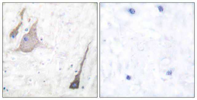
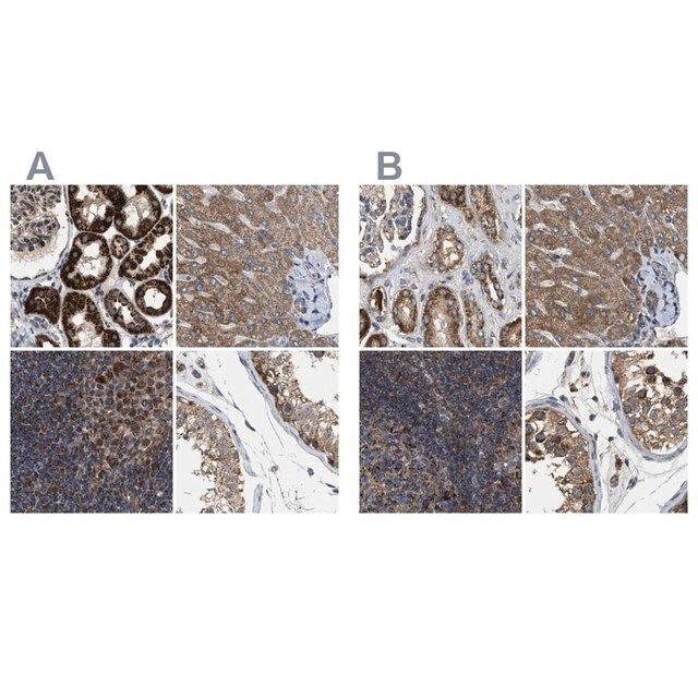
Immunohistochemistry Analysis: A 1:50 dilution from a representative lot detected Synapsin-1 in human, mouse, and rat cerebral cortex tissues.
Western Blotting Analysis: A representative lot detected synapsin Ia/Ib in mouse cortical neuron lysates (Medrihan, L., et al. (2013). Nat. Commun. 4:1512).
Western Blotting Analysis: A representative lot detected purified bovine brain synapsin Ia/Ib (Messa, M., et al. (2010). J. Cell Sci. 123(13):2256-2265).
Western Blotting Analysis: A representative lot detected synapsin-1 in Torpedo (electric ray) synaptosomal preparations and in GST-cyclophilin B pull-downs (Lane-Guermonprez, L., et al. (2005). J. Neurochem. 93(6):1401-1411).
Western Blotting Analysis: A representative lot detected synapsin-1 in the same rat brain subcellular fractions as cyclophilin B (Lane-Guermonprez, L., et al. (2005). J. Neurochem. 93(6):1401-1411).
Western Blotting Analysis: A representative lot detected synapsin Ia/Ib, but not IIa/IIb, in rat brain post-nuclear supernatants (Vaccaro, P., et al. (1997). Brain Res. Mol. Brain Res. 52(1):1-16).
Immunoprecipitation Analysis: A representative lot immunoprecipitated synapsin Ia/Ib, but not IIa/IIb, from rat brain synaptosomal preparations using protein G beads with pre-bound rabbit anti-mouse IgG (Vaccaro, P., et al. (1997). Brain Res. Mol. Brain Res. 52(1):1-16).
Immunofluorescence Analysis: Clone 10.22 ascites fluid was employed to localize synapsin-1 immunoreactivity within retinal inner plexiform layer (IPL) of floating or whole-mount rat retinas (Mandell, J.W., et al. (1992). J. Neurosci. 12(5):1736-1749).
Note: Clone 10.22 does not bind significantly to protein G. For immunoprecipitation application, pre-coat protein G beads with an anti-mouse IgG antibody is recommended (Vaccaro, P., et al. (1997). Brain Res. Mol. Brain Res. 52(1):1-16).
Research Sub Category
Developmental Neuroscience
质量
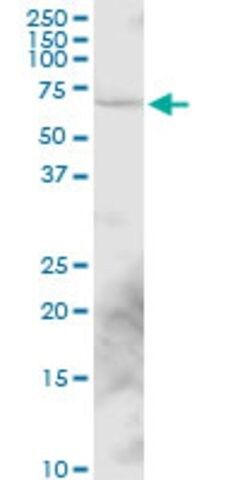
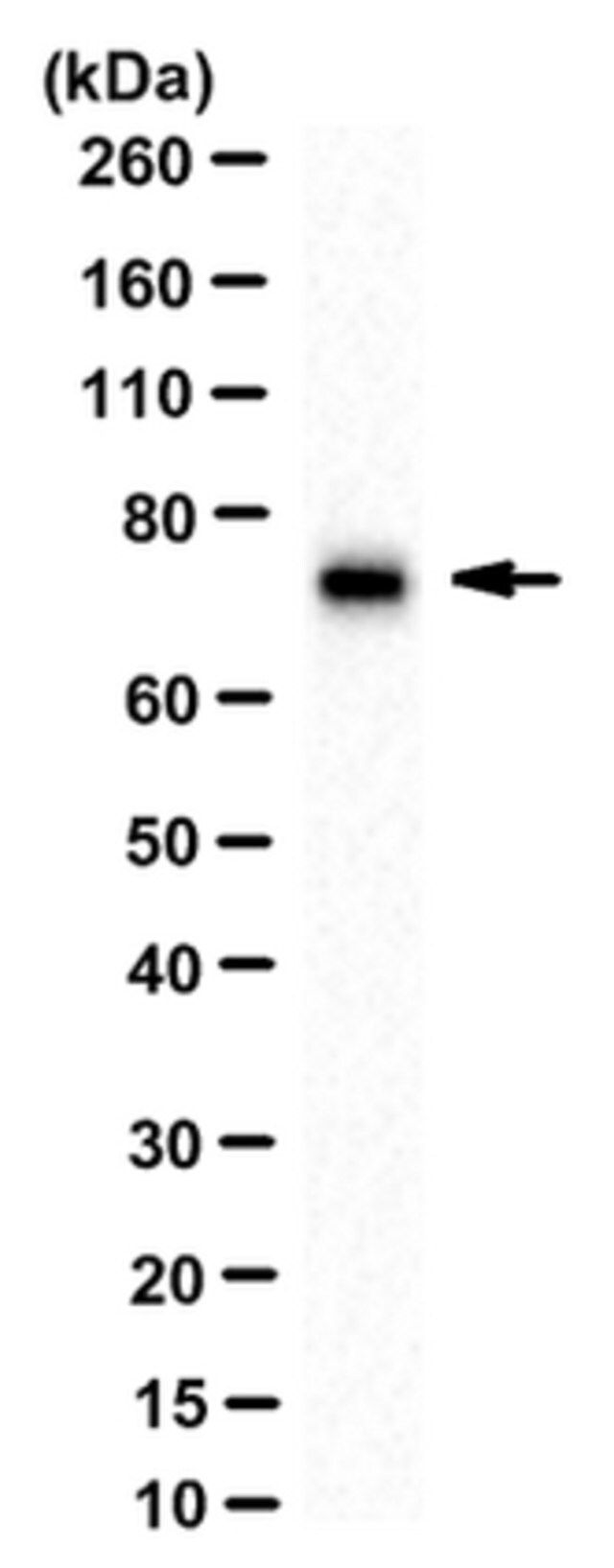
Evaluated by Western Blotting in rat brain cytosol tissue lysate.
Western Blotting Analysis: 0.5 µg/mL of this antibody detected Synapsin-1 in 10 µg of rat brain cytosol tissue lysate.
目标描述
~75 kDa observed.
外形
Format: Purified
Purified mouse monoclonal IgG1 antibody in buffer containing 0.1 M Tris-Glycine (pH 7.4), 150 mM NaCl with 0.05% sodium azide.
Protein G Purified
储存及稳定性
Stable for 1 year at 2-8°C from date of receipt.
其他说明
Concentration: Please refer to lot specific datasheet.
免责声明
Unless otherwise stated in our catalog or other company documentation accompanying the product(s), our products are intended for research use only and are not to be used for any other purpose, which includes but is not limited to, unauthorized commercial uses, in vitro diagnostic uses, ex vivo or in vivo therapeutic uses or any type of consumption or application to humans or animals.
基本信息
| eCl@ss | 32160702 |
| NACRES | NA.41 |
产品性质
| 质量水平 | 100 |
| 生物来源 | mouse |
| 抗体形式 | purified antibody |
| antibody product type | primary antibodies |
| 克隆 | 10.22, monoclonal |
| species reactivity | fish, human, bovine, rat, mouse |
| technique(s) | immunofluorescence: suitable immunohistochemistry: suitable (paraffin) immunoprecipitation (IP): suitable western blot: suitable |
| 同位素/亚型 | IgG1 |
| NCBI登记号 | NP_776616.1 |
| UniProt登记号 | P17599 |
| 运输 | wet ice |
| Gene Information | human ... SYN1(6853) |
安全信息
| 储存分类代码 | 12 - Non Combustible Liquids |
| WGK | WGK 1 |
| 闪点(F) | Not applicable |
| 闪点(C) | Not applicable |
Sigma-Aldrich

 m.cnreagent.com