

产品说明
一般描述
ZooMAb® antibodies represent an entirely new generation of recombinant monoclonal antibodies.Each ZooMAb® antibody is manufactured using our proprietary recombinant expression system, purified to homogeneity, and precisely dispensed to produce robust and highly reproducible lot-to-lot consistency. Only top-performing clones are released for use by researchers. Each antibody is validated for high specificity and affinity across multiple applications, including its most commonly used application. ZooMAb® antibodies are reliably available and ready to ship when you need them.
特异性
Clone 131-2A is a Mouse recombinant monoclonal antibody that detects fusion protein in Respiratory syncytial virus (RSV).
免疫原
A2 Respiratory Syncytial Virus (RSV) extract.
应用
Quality Control Testing
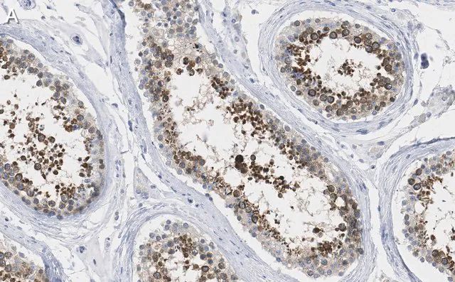
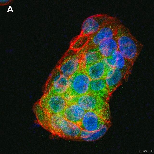
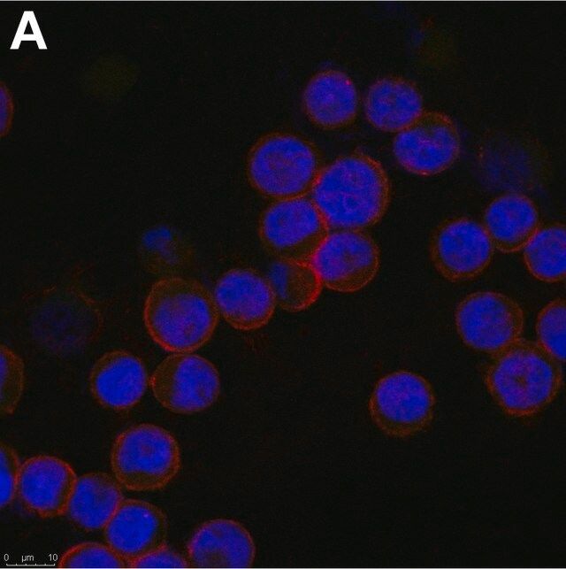
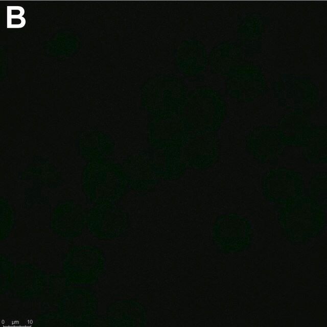
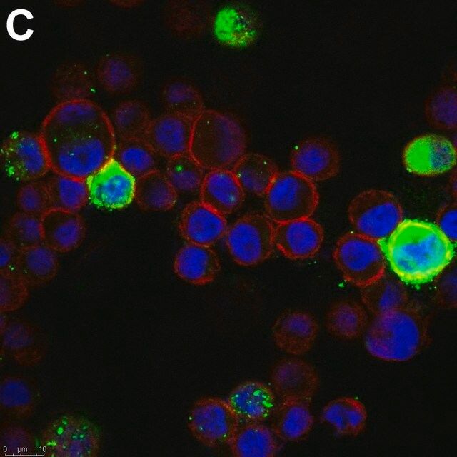
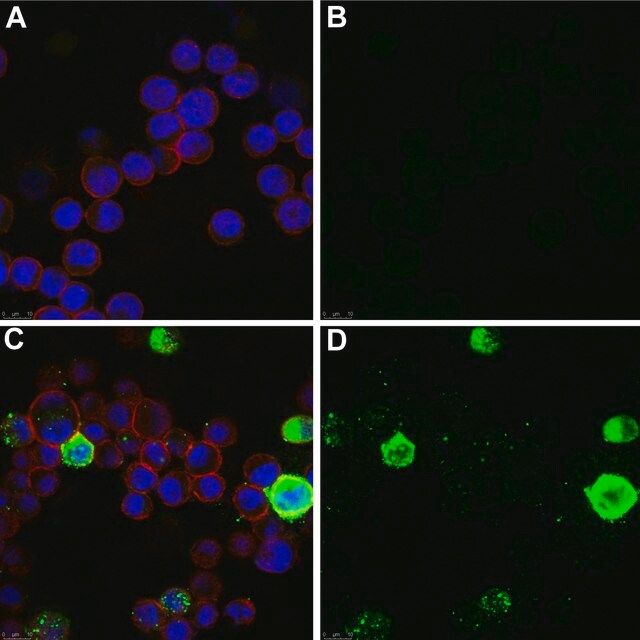
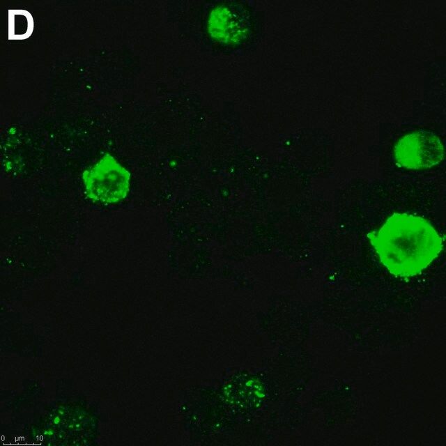
Evaluated by Immunocytochemistry in RSV infected HEp-2 cells.
Immunocytochemistry Analysis (ICC): A 1:1,000 dilution of this antibody detected RSV Fusion Protein in RSV infected HEp-2 cells.
Tested applications
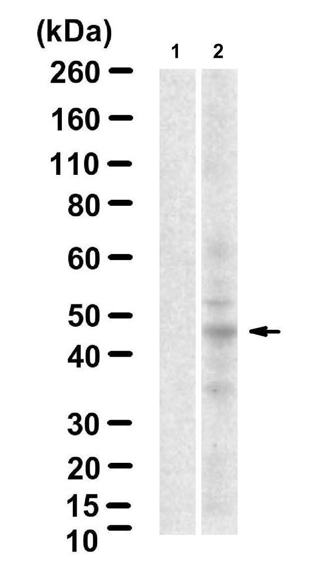
Western Blotting Analysis: A 1:1,000 dilution from a representative lot detected RSV Fusion Protein in lysate from RSV infected HEp-2 cells.
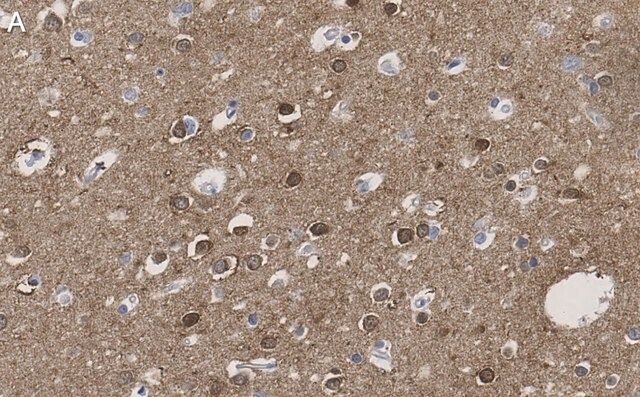
Immunohistochemistry (Paraffin) Analysis: A 1:1,000 dilution from a representative lot detected RSV Fusion Protein in RSV infected HEp-2 cells.
ELISA Analysis: A representative lot detected RSV Fusion Protein in ELISA applications (Newby, J., et. al. (2017). Nat Commun. 8(1):833).
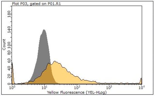
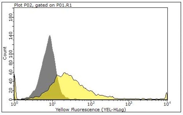
Flow Cytometry Analysis: 1 μg from a representative lot detected RSV Fusion Protein in one million RSV infected HEp-2 cells.
Flow Cytometry Analysis: A representative lot detected RSV Fusion Protein in Flow Cytometry applications (Preugschas, H.F., et. al. (2019). Cell Microbiol. 21(1):e12955).
Note: Actual optimal working dilutions must be determined by end user as specimens, and experimental conditions may vary with the end user
目标描述
Fusion glycoprotein F0 (UniProt: P03420) is encoded by the F gene (Gene ID: 1494475) in Human respiratory syncytial virus A (strain A2). Human respiratory syncytial virus (hRSV) is a pneumovirus that causes significant respiratory disease in premature and full-term infants. Fusion glycoprotein F0 of RSV is synthesized with a signal peptide (aa 1-15), which is subsequently cleaved off. The inactive F0 precursor is then cleaved at two sites (aa 109-110 and 136-137) by furin-like protease to give rise to produce mature F1 and F2 fusion glycoproteins. Fusion glycoprotein F1 is a single-pass type I membrane protein that can exist as homotrimer or can form disulfide-linked heterodimer with fusion glycoprotein F2. Its N-terminus has a hydrophobic fusion peptide that inserts into the target host membrane. It is buried in the center of the trimer cavity before cleavage by host furin. The fusion glycoprotein F2 is the major determinant of the species specificity of RSV infection. Fusion protein can exist in at least 3 conformational states: pre-fusion native state, pre-hairpin intermediate state, and post-fusion hairpin state. During viral and plasma cell membrane fusion, the coiled coil regions assume a trimer-of-hairpins structure, positioning the fusion peptide in close proximity to the C-terminal region of the ectodomain. This fusion is pH independent and occurs at the plasma or endosomal membrane. The fusion protein is reported to be involved in the entry into the host cell through the interaction with host IGF-R1. This interaction activates protein kinase z that recruits host NCL/nucleolin to the apical cell surface where it can bind fusion glycoprotein F1. Later in infection, fusion protein expressed at the plasma membrane of infected cells can mediate fusion with adjacent cells to form syncytia, a cytopathic effect that could lead to tissue necrosis. Fusion protein can also trigger p53-dependent apoptosis in cells. This ZooMAb® recombinant monoclonal antibody, generated by our propriety technology, offers significantly enhanced specificity, affinity, reproducibility, and stability over conventional monoclonals. (Ref.: McLellan, JS., et al. (2013). Science. 340(6136); 1113-1117; Olivier, A., et al. (2009). Int. J. Exp. Pathol. 90(4); 431-438; Schlender, J., et al. (2003). J. Virol. 77(8); 4609-4616).
外形
Purified recombinant mouse monoclonal antibody IgG, lyophilized in PBS, 5% Trehalose, normal appearance a coarse or translucent resin. The PBS/trehalose components in the ZooMAb formulation can have the appearance of a semi-solid (bead like gel) after lyophilization. This is a normal phenomenon. Please follow the recommended reconstitution procedure in the data sheet to dissolve the semi-solid, bead-like, gel-appearing material. The resulting antibody solution is completely stable and functional as proven by full functional testing. Contains no biocide or preservatives, such as azide, or any animal by-products. Larger pack sizes provided as multiples of 25 μL.
重悬
300 μg/mL after reconstitution at 25 μL per vial. Please refer to guidance on suggested starting dilutions and/or titers per application and sample type.
储存及稳定性
Recommend storage of lyophilized product at 2-8℃; Before reconstitution, micro-centrifuge vials briefly to spin down material to bottom of the vial; Reconstitute each vial by adding 25 μL of filtered lab grade water or PBS; Reconstituted antibodies can be stored at 2-8℃, or -20℃ for long term storage. Avoid repeated freeze-thaws.
法律信息
ZooMAb is a registered trademark of Merck KGaA, Darmstadt, Germany
免责声明
Unless otherwise stated in our catalog or other company documentation accompanying the product(s), our products are intended for research use only and are not to be used for any other purpose, which includes but is not limited to, unauthorized commercial uses, in vitro diagnostic uses, ex vivo or in vivo therapeutic uses or any type of consumption or application to humans or animals.
产品性质
| 质量水平 | 200 |
| 生物来源 | mouse |
| 重组 | expressed in HEK 293 cells |
| 偶联物 | unconjugated |
| 抗体形式 | purified antibody |
| antibody product type | primary antibodies |
| 克隆 | 131-2A, recombinant monoclonal |
| 描述 | recombinant, expressed in HEK 293 cells |
| 产品线 | ZooMAb® learn more |
| 形式 | lyophilized |
| 分子量 | calculated mol wt 63.45 kDa observed mol wt ~45 kDa |
| 纯化方式 | using protein G |
| species reactivity | virus |
| 包装 | antibody small pack of 25 μL |
| 环保替代产品特性 | Waste Prevention Designing Safer Chemicals Design for Energy Efficiency Learn more about the Principles of Green Chemistry. |
| 增强验证 | recombinant expression Learn more about Antibody Enhanced Validation |
| technique(s) | ELISA: suitable flow cytometry: suitable immunocytochemistry: suitable immunohistochemistry (formalin-fixed, paraffin-embedded sections): suitable western blot: suitable |
| 同位素/亚型 | IgG2aκ |
| 表位序列 | Unknown |
| Protein ID登记号 | QEI22738.1 |
| UniProt登记号 | P03420 |
| 环保替代产品分类 | Aligned |
| 运输 | ambient |
| 储存温度 | 2-8℃ |
安全信息
| 储存分类代码 | 13 - Non Combustible Solids |
| WGK | WGK 1 |
| 闪点(F) | Not applicable |
| 闪点(C) | Not applicable |
Sigma-Aldrich

 m.cnreagent.com